

Abstract
Introduction. T-cell large granular lymphocytosis (T-LGL) is a low grade lymphoproliferative disorder, often clinically manifest as bone marrow failure. Treatment with immunosuppressive therapies is effective, but the dominant clone may persist even in responding patients. The pathogenesis of T-LGL has not been fully elucidated. In this study, we performed single cell RNA sequencing (sc-RNA seq) and V(D)J profiling to discern clonotypes and gene expression patterns of T lymphocytes from T-LGL patients who were sampled before and after treatment.
Methods. Blood was obtained from patients participating in a phase 2 protocol of alemtuzumab as second line therapy (NCT00345345; Dumitriu B et al, Lancet Haematol 2016). Leukapheresis was performed in 13 patients (M/F 7/6; median age 51 years, range 26-85) before and after 3-6 months alemtuzumab administration and in 7 age-matched healthy donors. Cryopreserved blood was enriched for T cells with the EasySep Human T cell Isolation Kit (Stem cell). sc-RNA seq was performed on the 10XGenomics Chromium Single Cell V(D)J + 5' Gene Expression platform, and sequencing obtained on the HiSeq3000 Platform. Barcode assignment, alignment, unique molecular index counting and T cell receptor sequence assembly were performed using Cell Ranger 2.1.1.
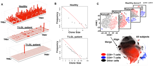
Results. Four hundred fifty thousand cells from 13 patients and 107,000 cells from 7 healthy donors were profiled. We measured productive TCR chains (which fully span the V and J regions, with a recognizable start codon in the V region and lacking a stop codon in the V-J region, thus potentially generating a protein). We detected at least one productive TCR α-chain in 50%, one productive TCR β-chain in 69% and paired productive αβ-chains in 47% of all cells. There was loss of TCR repertoire diversity in patients which was quantified by Simpson's diversity index; most patients showed oligoclonal or, less frequently, monoclonal expansion of the TCR repertoire (Fig. A). Regardless of clinical response, alemtuzumab treatment did not correct the low TCR repertoire diversity. TCR repertoires can be classified as "public", when they express identical TCR sequences across multiple individuals, or "private", when each individual displays distinct TCR clonotypes. No TCRA or TCRB CDR3 homology among patients was observed: most TCR clonotypes appeared to be private. Our data suggests that T-LGL is etiologically heterogenous disease, consistent with T cell expansion in response to a variety antigens, in diverse HLA contexts, or randomly. Despite differences of TCR among patients and healthy donors, and the presence of large clones in patients, distribution of TCR diversity followed the power law distribution in healthy donors and patients (Fig. B, showing the negative linear relationship between logarithmic expression of clone frequency and clone size). The observed distribution is consistent with a somatic evolution model, in which cell fitness depends on cellular receptor response to specific antigens and stimulation of cells by cytokine and other signals from the environment; fitted clones have higher birth-death ratios and thus expand (Desponds J et al, PNAS 2016).
CD4 and CD8 T cells can be virtually separated by imputation from their transcriptomes (Fig. C). Comparison of gene expression between patients and healthy donors showed dysregulation of genes involved in pathways related to the immune response and cell apoptosis, consistent with a pathophysiology of T cell clonal expansion. We used diffusion mapping, which localizes datapoints to their eigen components in low-dimesional space, to characterize sources contributing to the gene expression phenotype: the first component was mainly from T cell activation and the second was associated with TCR expression. In LGL the T cell transcriptome appeared to be shaped by both lineage development and TCR rearrangement.
Conclusion. We describe at the single cell level T clonal expansion profiles in T-LGL, pre- and post-treatment. Single cell analysis allows accurate recovery of paired α and β chains in the same cell and demonstrates a continuum of cell lineage differentiation. We found a range of differences in transcriptome and TCR repertoires across patients. Transcriptome data, coupled with detailed TCR-based lineage information, provides a rich resource for understanding of the pathology of T-LGL and has implications for prognosis, treatment, and monitoring in the clinic.
Young:GlaxoSmithKline: Research Funding; CRADA with Novartis: Research Funding; National Institute of Health: Research Funding.

This feature is available to Subscribers Only
Sign In or Create an Account Close Modal